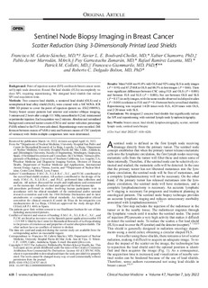
Sentinel Node Biopsy Imaging in Breast Cancer Scatter Reduction Using 3-Dimensionally Printed Lead Shields
| dc.rights.license | Attribution-NonCommercial-NoDerivatives 4.0 International | * |
| dc.contributor.author | chamorro, xabier | |
| dc.contributor.other | Canete-Sanchez, Francisco M. | |
| dc.contributor.other | Boulvard-Chollet, Xavier L. E. | |
| dc.contributor.other | Marrodan, Pablo Javier | |
| dc.contributor.other | Garrastachu Zumaran, Puy | |
| dc.contributor.other | Ramirez Lasanta, Rafael | |
| dc.contributor.other | Colletti, Patrick M. | |
| dc.contributor.other | Giammarile, Francesco | |
| dc.contributor.other | Delgado Bolton, Roberto C. | |
| dc.date.accessioned | 2022-11-10T14:38:44Z | |
| dc.date.available | 2022-11-10T14:38:44Z | |
| dc.date.issued | 2022 | |
| dc.identifier.issn | 1536-0229 | en |
| dc.identifier.other | https://katalogoa.mondragon.edu/janium-bin/janium_login_opac.pl?find&ficha_no=168050 | en |
| dc.identifier.uri | https://hdl.handle.net/20.500.11984/5831 | |
| dc.description.abstract | Background Point of injection scatter (SPI) confounds breast cancer sentinel lymph node detection. Round flat lead shields (FLSs) incompletely reduce SPI, requiring repositioning. We designed lead shields that reduce SPI and acquisition time. Methods Two concave lead shields, a semioval lead shield (OLS) and a semispherical lead alloy shield (SLS), were created with a SICNOVA JCR 1000 3D printer to cover the point of injection (patent no. ES1219895U). Twenty breast cancer patients had anterior and anterior oblique imaging, 5 minutes and 2 hours after a single 111 MBq nanocolloid in 0.2 mL intratumoral or periareolar injection. Each acquisition was 2 minutes. Absolute and normalized background corrected scatter counts (CSCs) and scatter reduction percentage (%SR) related to the FLS were calculated. Repositionings were recorded. Differences between means of %SR (t test) and between means of CSC (analysis of variance) with Holm multiple comparison tests were determined. Results Mean %SR was 91.8% with OLS and 92% using SLS in early images (P = 0.91) and 87.2%SR in OLS and 88.5% in late images (P = 0.66). There were significant differences between CSC using FLS and OLS (P < 0.001) and between FLS and SLS (P < 0.001), but not between OLS and SLS (P = 0.17) in early images, with the same results observed in delayed studies (P < 0.001 in relation to FLS and P = 0.1 between both curved lead shields). Repositioning was required 14/20 times with FLS, 4/20 times with OLS, and 2/20 times with SLS. Conclusions We designed 2 concave lead shields that significantly reduce the SPI and repositioning with sentinel lymph node lymphoscintigraphy. | en |
| dc.language.iso | eng | en |
| dc.publisher | Wolters Kluwer | en |
| dc.rights | © 2022 The Authors | en |
| dc.rights.uri | http://creativecommons.org/licenses/by-nc-nd/4.0/ | * |
| dc.subject | Breast cancer | en |
| dc.subject | lead shield | en |
| dc.subject | lymphoscintigraphy | en |
| dc.subject | scatter | en |
| dc.subject | sentinel lymph node | en |
| dc.subject | sentinel node biopsy | en |
| dc.title | Sentinel Node Biopsy Imaging in Breast Cancer Scatter Reduction Using 3-Dimensionally Printed Lead Shields | en |
| dcterms.accessRights | http://purl.org/coar/access_right/c_abf2 | en |
| dcterms.source | Clinical Nuclear Medicine | en |
| local.description.peerreviewed | true | en |
| local.description.publicationfirstpage | 618 | en |
| local.description.publicationlastpage | 624 | en |
| local.identifier.doi | https://doi.org/10.1097/RLU.0000000000004274 | en |
| local.contributor.otherinstitution | https://ror.org/040xzg562 | es |
| local.contributor.otherinstitution | VietTin Land Investment and Consultant | en |
| local.contributor.otherinstitution | https://ror.org/03taz7m60 | en |
| local.contributor.otherinstitution | https://ror.org/02zt1gg83 | en |
| local.contributor.otherinstitution | https://ror.org/01cmnjq37 | fr |
| local.source.details | Vol. 47. N. 7. P.p. 618-624, | en |
| oaire.format.mimetype | application/pdf | |
| oaire.file | $DSPACE\assetstore | |
| oaire.resourceType | http://purl.org/coar/resource_type/c_6501 | en |
| oaire.version | http://purl.org/coar/version/c_970fb48d4fbd8a85 | en |
Item honetako fitxategiak
Item hau honako bilduma honetan/hauetan agertzen da

Bestelakorik adierazi ezean, itemaren baimena horrela deskribatzen da: Attribution-NonCommercial-NoDerivatives 4.0 International